 |
||
|
|
||
|
 Partners:   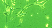 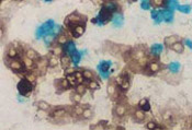
|
Confocal | Phase Contrast | Electron Microscopy | Cell Culture | Fractionation The discovery and study of cells was dependent on the development of the microscope. Further refinements and adaptations have allowed study of subcellular components at the molecular level. Here, we review some of the common methods used to study cells and tissues. Under the standard light microscope thin sections of tissues are examined by transillumination. Light is passed through a condenser that collects the light into a focused beam that then passes through the specimen and is collected by an objective lens that magnifies the image (anywhere from 4X to 200X). The specimen is viewed through an ocular lens that also magnifies the image (usually 4X). The total magnification is obtained by multiplying the magnifying power of the objective and ocular lenses. Image quality is dependent on the resolving power of the microscope, which is largely determined by the quality of the objective lens. The light microscope has a limiting resolution of about 0.1 micron (objects smaller than this cannot be resolved) which allows a magnification of over 1000X without a loss of quality. Often a camera is attached to the microscope via a separate port so that pictures can be taken while viewing through the ocular. Very sensitive video cameras are used to capture and transfer digital images to computer programs for image analysis. Most cells and tissues in their natural state contain little pigment that would absorb light. Therefore, they are normally translucent to transmitted light and little detail can be seen. Consequently, methods have been developed to stain cells, subcellular components and tissue components and structures. Most classical stains require a fixed or preserved specimen. Preservation of the specimen is necessary to preserve cellular structures in a "natural" state against intracellular digestion and desiccation. When tissues or cells must be thinly sectioned, the aqueous environment is replaced by a resin or embedding media such as paraffin or epoxy that penetrates the cell and adds rigidity to the tissue. These embedding media are usually not hydrophillic and sometimes lipids are removed. To limit the loss of lipids or study enzyme activity, sometimes the tissues are cut when frozen rather than fixing and embedding them. In the ideal tissue preparation the tissue retains the same structure and molecular composition as the living tissue. This is very rare and usually artifacts from the preparation process are present. Phase Contrast Microscopy can be used to image transparent cells. The principle behind this type of microscopy is that light changes speed and direction when passing through cellular and extracellular structures of different refractive indices. This causes some structures to appear lighter or darker relative to each other. A modification of this is Normarski Differential Interference Microscopy that uses polarized light and gives a three dimensional look at transparent objects. Confocal microscopy uses lasers and computers to produce three-dimensional images of living cells and tissue slices. Optical sections of the specimen are collected by laser scanning and stored in the computer. The information from each visual plane can be viewed and reconstructed into three dimensional projections of the specimen. Fluorescence Microscopy utilizes substances (fluorophores) that, when irradiated by a certain wavelength of light emit light of a longer wavelength. Tissue sections are usually irradiated with ultraviolet light so that the emission is in the visible portion of the spectrum. The microscope used has a strong ultraviolet light source and special filters that eliminate ultraviolet light before it gets to the observers eyes. Some fluorescent compounds have an affinity for nucleic acids allowing the localization of nucleic acids with cells. Others are specific for the mitochondria. Fluorescent probes can be made by chemically coupling the fluorophore to a relevant molecule such as a hormone or antibody. Electron Microscopy is based on the interaction of electrons with tissue components and electron dense stains. The electron microscope is capable of 0.1 nanometer resolution. Magnifications can be as much as 400 times greater than those achieved in light microscopy. The principle behind transmission electron microscopy or TEM is that an electron beam is passed through a stained ultrathin section of a tissue that has been embedded in plastic. The thin sections are cut with diamond or glass knives and mounted on metal grids prior to staining. The electron dense stain (lead or uranium) is differentially taken up by the cellular structures and therefore the electrons are absorbed more by some structures than others. The electrons that pass through the specimen fall on a photographic film or digital image plate to form an image. Scanning electron microscope permits pseudo-three-dimensional views of the surface of specimens. Specimens are covered with an electron dense material and a narrow electron beam in passed over the surface. At each point, the electron beam produces emitted electrons from the metal coating of the specimen. A detector captures these emitted electrons and the brightness of a synchronous cathode ray tube is modified to produce a three dimensional image that has highlights and shadows. Cell and tissue culture allows for the direct study of cell behavior in vitro. Chemically defined media, growth factors, hormones and serum components simulate the normal environment in a culture dish. Infectious agents such as protozoa and viruses that grow only within cells can be studied using cell culture techniques. Cells are collected by enzymatic or mechanical disruption of tissues and isolated to culture media as suspension or contact dependent cultures. Normal cells have finite, genetically programmed life span. However, transformed cells such as those found in cancer, become immortal cell lines and can go through many more cell divisions and have a much longer life. Cells can be harvested and frozen in liquid nitrogen for later reconstitution and use in cell culture experiments. Separation of cellular components from one another is accomplished by differential centrifugation. Centrifugal force is used to separate organelles and cellular components as a function of their sedimentation coefficients. The coefficient is based on the size, form, and density of the particle. Cells are mechanically lysed and the homogenate is subjected to successive centrifugation at increasing speeds. At each step the resultant pellet contains a different cellular component and the supernatant is collected for the next step. For instance, the first step might pellet the nuclei leaving the other cellular components in the supernatant. After the second step mitochondria and lysosomes are collected in the pellet and the supernatant subjected to the next spin. The size and density of the cellular components in the pellet decreases with each step in the process. Cell fractionation allows detailed study of cellular components obtained in a relatively pure state. Immunocytochemical methods allow the study of the presence and activity of specific macromolecules in cells and tissues. Antigen-antibody, and receptor-hormone interactions are exploited by the labeling of proteins with fluorescent molecules, enzymes or electron dense molecules. These labels make the molecule visible in the microscope without causing a loss of the protein's biologic activity. Labeled antibodies or hormones bind only to their antigens or receptors, respectively, thereby permitting localization of specific antigens in tissue specimens. In direct immunocytochemistry, an antibody is made specific to the antigen. The more sensitive indirect approach uses an unlabeled primary antibody to the antigen and a labeled secondary antibody that binds to the primary antibody and makes the complex visible under the microscope.
|
|
|
|
||